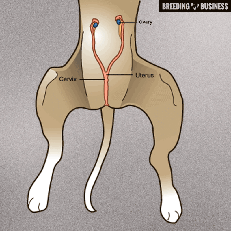
The canine uterus anatomy model is a high quality and detailed education tool and includes a display stand, teacher guide and student worksheet. A female dog’s reproductive system involves the uterus, the cervix, the oviducts, the ovaries, and the vagina.
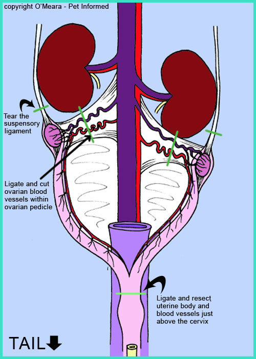
Spaying a Female Dog — Procedures, Risks, Benefits
The paired ovaries are the female gonads;

Dog uterus anatomy. The uterine horns extend from each ovary and join to form the body of the uterus. The vagina is very long relative to that in other species. The minor duodenal papilla, which receives the accessory pancreatic duct, is generally present in the dog but present in only a minority (20%) of cats.
This is what’s responsible for the tie that occurs during mating. A pregnant female dog's anatomy involves the two ovaries which produce eggs, cervix, fallopian tubes, and the uterus. The most common smooth muscle tumor of the uterus is what is classified as leiomyoma.
During copulation, the bulbus glandis engorges with blood which makes it swell. These variations occur in both the anatomical types of uterus as well as the uterine horn appearance and endometrial linings. A dog's body will change as she goes through heat cycles, however, her basic anatomy remains unchanged.
Female vs male dog anatomy male dog anatomy. The oviducts are small ducts that transport the oocytes from the ovary to the uterus. The eggs travel from the ovaries to her oviducts, where the eggs are fertilized by sperm.
Ejaculation will occur and sperm cells will enter the uterus and make their way to the oviduct, where their union with the. The most common neoplasm of the wall of the uterus in the dog is the smooth muscle tumor (leiomyoma). In the dog, a tie usually occurs in which the male and female are held together physically, with the vagina tightly enclosed around the glans penis.
Not only are females generally smaller than males in the dog world; The parts (body) of the uterus (in female dog) other organs and structures related to the male and female dogs (will enlist in the figure) now, this is time to learn details of these organs and structures from the dog pelvis. When the female is pregnant, the fetuses are arranged in a row in both horns.
232 the body of the uterus is located in the pelvic and abdominal cavities; When the neoplasm has a abundant fibrous tissue with the smooth muscle, they are called fibroleiomyoma. The uterus is the reproductive organ with the most species variations.
From midvagina cranially, the vaginal vault is decreased in size significantly by the presence of the dorsal median postcervical fold, a piece of tissue that hangs from the dorsal surface, or ceiling, of the vagina. They also play a very important role in sustaining the pregnancy. The ovaries (oviducts), the uterus, the cervix, and;
Anatomy is a branch of biology and medicine that studies the morphology and structure of living organisms. The detailed structure depends on a lot of factors such as the dog breed, age, and weight. They contain the oocytes and are responsible for much of the dog's reproductive hormone production.
99 cat uteri are usually 3 to 4 mm wide. The male has what’s known as a bulbus glandis. The uterine horns during the seventh week were.
Duplex uteri are seen in rabbits and marsupials. The minor papilla is absent when the accessory pancreatic duct has atrophied. The uterus becomes the womb for her puppies during their gestation period.
They have a few other differences too. The female dog anatomy external organ is the vulva which opens to the vagina. The nonpregnant uterine size varies considerably among species but, more importantly, with previous pregnancies, stage of estrus cycle, and age.
To understand the difference, a basic understanding of dog reproductive system and it’s anatomy is needed. The dog’s pelvic bones include the pelvic girdles, thigh, leg, and hind paw. The female dog’s reproductive anatomy consists of:
The uterine horns during the first week postpartum were dilated and edematous. 2021 ultimate guide to dog anatomy. The ovaries are the organs that are responsible for the production of unfertilized eggs in the female.
— union of a haploid oocyte and a haploid spermatozoon, producing a diploid zygote The anatomy of the vagina of the dog is complex. Almost 60 days in horse, cattle, human).
Endometrium bartel et al (2014) studied the lipid laden surface endometrial cells of the uterus in metestrus/diestrus using electron microscopy and immunohistochemistry. The fertilized eggs continue to the uterus to mature, where they develop into embryos, attached to the uterine wall by a placenta. The canine reproductive system for females differs greatly from the male counterpart.
The normal canine uterus is a bicorniate structure composed of 2 horns and a body. Urethra and bladder in the excretion of liquid wastes and the reproductive system that includes the female uterus, ovaries, fallopian tubes and vagina and the male testes, epididymis, vas deferens and penis. The suspensory ligament of the ovarytethers the ovary to the dorsal abdominal wall.
Completely separate uterine horns each with their own cervical canal. The long arms of the uterus are called the horns, and the short stem is called the body. Whereas a male may be stimulated at any time, a female must be in a designated heat period for copulation to flourish successfully.
And female dog anatomy aims at making a study of all parts of the female dog’s body. Of organ development (about 30 days in dog, cat, sheep, pig; This results in accumulation of pus in the uterus of sexually intact bitches, typically occurring between 4 weeks and 4 months after a heat cycle.

Pyometra in Dogs uterus infection, pus, causes, signs
Dog Spaying Surgery Everything you need to know about
LORI.canine

A dogs uterus isnt saclike as it is in humans but it has

Neutering And Spaying Dogs, What Are The Risks